Fetal Echocardiography
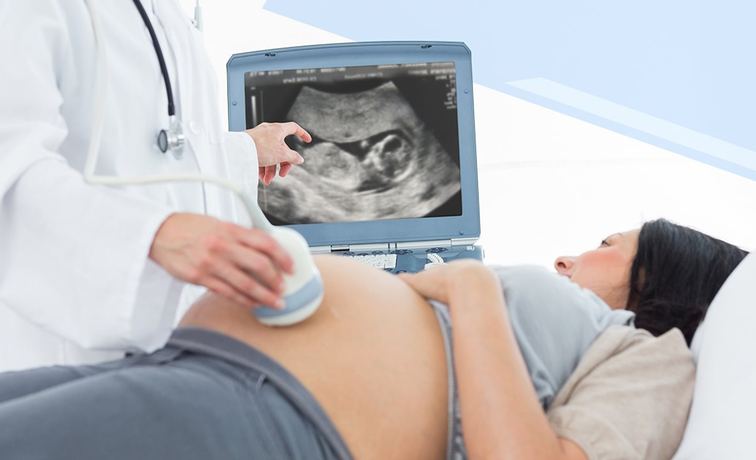
Fetal Echocardiography is an advanced ultrasound test that focuses on the structure and function of a baby’s heart while still in the womb. Performed between 18 and 24 weeks of pregnancy, this scan helps detect congenital heart defects (CHDs) and other cardiac conditions early, allowing timely care and intervention.
When is Fetal Echocardiography Recommended?
- Family history of heart defects
- Abnormal results from routine ultrasound
- Diabetes or autoimmune conditions in the mother
- Use of certain medications during pregnancy
- IVF or multiple pregnancies
Using high-resolution ultrasound and Doppler imaging, our specialists examine the baby’s heart chambers, valves, blood flow, and rhythm to detect abnormalities in structure or function.
Why Choose Our Center?
- Performed by expert fetal cardiologists and radiologists
- Latest imaging technology for accurate diagnosis
- Safe, non-invasive, and painless procedure
- Comprehensive report with guidance for follow-up care
- Coordination with pediatric cardiologists, if required
Fetal Echocardiography provides essential information that helps prepare for a baby’s care after birth, especially if any heart condition is identified. Our expert team ensures compassionate care and expert diagnostics for a healthy start to your baby's life.